16 Nov 2015
Investigating upper airway obstruction in horses

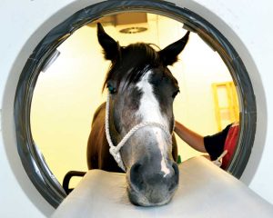
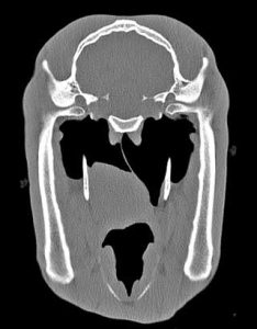
Figure 7b. Horse’s throat in CT “doughnut”.
Upper airway obstruction is common in horses and of major consequence to equine athletes in all disciplines.
Major advances in technology have created an array of useful imaging techniques available to the equine clinician investigating the equine pharynx and larynx, which are now routinely employed in the diagnosis of equine upper airway obstruction.
However, a thorough clinical examination at rest and during appropriate exercise remains the cornerstone of any investigation.
Recognising and identifying the cause of an abnormal respiratory noise at exercise is an important skill to acquire, which takes practice and an understanding of the normal equine respiratory cycle during exercise. Limb movement and the respiratory cycle are intimately linked in horses moving at speed – typically in a 1:1 ratio.
It is essential to be able to differentiate between the inspiratory and expiratory phases of the respiratory cycle, which is most easily achieved by exercising the horse at a canter; expiration begins after the leading limb hits the ground.
Most normal horses make some expiratory sound, but inspiratory noises are characteristic of respiratory obstruction (many horses so affected will also make loud expiratory sounds as well).
Clinicians must also be aware correctly interpreting such sounds also requires sufficient hearing ability. In particular, the inspiratory “whistle” characteristically produced by horses suffering from recurrent laryngeal neuropathy (and some other causes of upper airway obstruction) is heard most effectively by the female ear and by men who are under 40 years of age.
Some pharyngeal and laryngeal conditions readily lend themselves to a diagnosis following a clinical evaluation, typically when combined with an endoscopic examination of the upper respiratory tract during resting respiration.
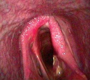
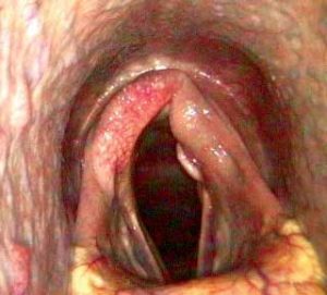
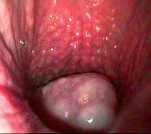
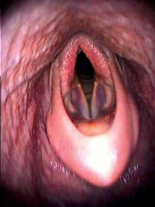
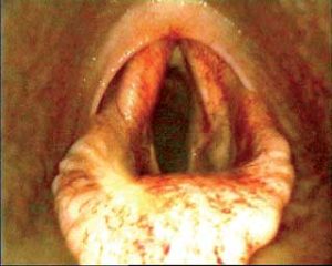
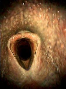
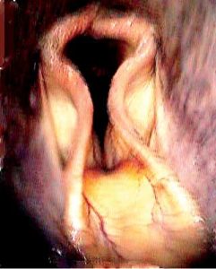
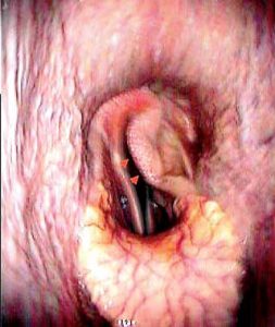
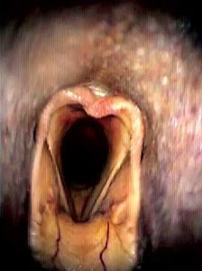
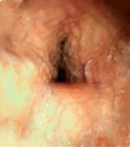
Examples of these conditions include the more severe stages of recurrent laryngeal neuropathy (laryngeal hemiplegia; Figure 1), arytenoid chondritis (Figure 2), swellings associated with the guttural pouches (that is, tympany or empyema; Figure 3), large subepiglottal cysts (Figure 4) and persistent epiglottal entrapment (Figure 5).
It is generally accepted assessing laryngeal symmetry is most satisfactorily achieved by an endoscopic examination carried out via the right nostril, as this obviates the appearance of artefactual “collapse” of the left side of the larynx caused by perspective distortion; the left arytenoid being typically affected by recurrent laryngeal neuropathy in the horse.
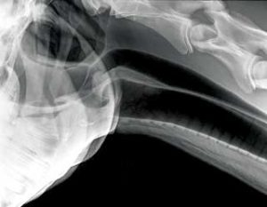
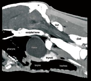
However, a number of conditions cannot be diagnosed by these means alone. In such cases, the use of radiography (Figures 6a and 6b), CT, diagnostic ultrasonography or dynamic endoscopy are often necessary to complete the investigation fully and to make an accurate diagnosis. These include those pharyngeal cysts not readily visible during routine endoscopy and some cases of fourth branchial arch anomaly.
In particular, the availability of CT for the standing patient (Figures 7a and 7b) has significantly enhanced the diagnosis of such conditions (Figures 8 and 9). This technique allows the creation of a 3D image of the throat by making radiographic “slices” of the area.
Without doubt, dynamic endoscopy in particular (Figure 10), has revolutionised the diagnosis of dynamic upper airway obstruction in performance horses of all types and permits the diagnosis of various causes of upper airway obstruction, which could not be identified by any other means – for example, intermittent dorsal displacement of the soft palate (Figure 11).
In the past, making a diagnosis of such conditions was largely a matter of educated guesswork. Its introduction and regular use in equine clinical practice has also allowed the demonstration of a number of novel conditions.
Examples of such previously unrecognised conditions include medial displacement of the aryepiglottal folds (Figure 12), dynamic laryngeal collapse (Figure 13), ventromedial displacement of the corniculate process of the arytenoid cartilage (Figure 14), nasopharyngeal collapse (Figure 15), epiglottal retroversion, cricotracheal ligament collapse and intermittent epiglottal entrapment.
It has also allowed more accurate identification of cases of recurrent laryngeal neuropathy, which are causing significant laryngeal obstruction at exercise.
Until relatively recently, the only means of performing dynamic endoscopy involved the use of a treadmill. This was potentially quite inconvenient as horses had to be acclimatised to exercising on the treadmill and some animals were reluctant to do so.
However, during the past decade, remote dynamic endoscopy using the so-called “over-ground endoscope” has transformed the process and horses can be examined during normal exercise.

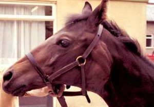
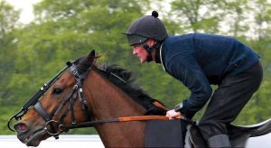
The equipment is completely portable and attached to the horse during exercise (Figure 16). It allows a video recording of the examination, which can be analysed after exercise, and a robust portable screen, held by the clinician during the examination, allows examination of the image by telemetry. This ensures the endoscope is positioned satisfactorily in the airway and also allows an instant diagnosis of the more obvious conditions.
Replay, sometimes using frame-by-frame analysis, is necessary to make a diagnosis with some of the more subtle or transient causes of upper airway obstruction.
Such improvements in diagnostic accuracy have not necessarily been matched by similar improvements in therapy, but these advances in imaging technology have at least reduced the use of inappropriate treatments based upon guesswork and misdiagnosis.
Undoubtedly, the more accurate diagnosis and critical evaluation of surgical treatment using dynamic diagnostic techniques will continue to increase our understanding of upper airway pathology and lead to more successful, evidence-based surgical and medical treatments. This is one of the main directions in which clinical research is heading.
However, in the absence of this sort of information, ensuring client satisfaction will continue to depend on managing expectations, scientific honesty and first-rate communication.
Acknowledgement
The author would like to acknowledge the assistance of Lorraine Palmer, Lewis Smith and Tim Barnett in obtaining some of the images used in this article.
Latest news


