11 Jan 2016
Joint pain in dogs related to osteoarthritis – part 3

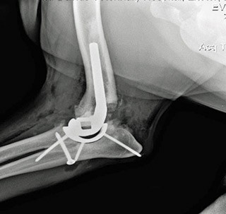
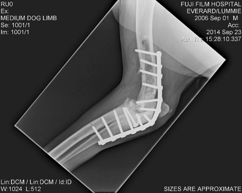
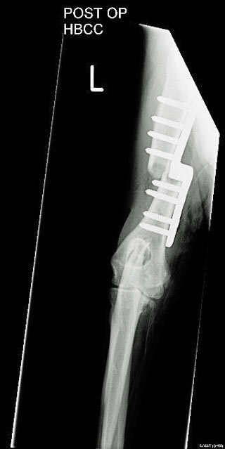
Figure 8. Postoperative image of an elbow arthrodesis.
Osteoarthritis (OA) is a common, painful condition of dogs that can be challenging to manage successfully in practice.

Numerous surgical options are available for management of OA and a full discussion of advantages and disadvantages should be held with owners of affected pets. Each case should be considered individually, based on the owners’ expectations, patient features and joint involved.
Options include joint replacement, excision arthroplasty, arthrodesis, bone realignment surgery (osteotomy) or arthroscopy with “joint debridement” procedures (Table 1).
Joint replacement surgery
Total joint replacement surgery is considered a standard treatment for advanced degenerative joint conditions in humans (Allen, 2012) and is becoming a readily available and increasingly affordable option for the treatment of advanced cases of hip, stifle and elbow osteoarthritis in dogs.
Customised joint replacements have also been reported for individual cases of painful shoulder and tibiotarsal joint conditions and will also inevitably become more available in the future (Sparrow et al, 2014).
When considering joint replacement, possible complications and costs must always be discussed fully with the owner. This conversation is best held between the surgeon performing the operation and the client during an initial joint replacement assessment consultation.
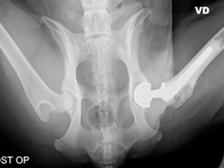
Total hip replacement (THR) is the preferred surgical option for the management of clinically significant hip OA in all breeds and sizes of dog – particularly where conservative treatment has failed. Excellent clinical results have been reported in a large number of clinical cases (Guerrero and Montavon, 2009; Liska, 2010; Lascelles et al, 2010; Marino et al, 2012). Following successful THR surgery, one expects a rapid and full return to normal joint function (Figure 1).
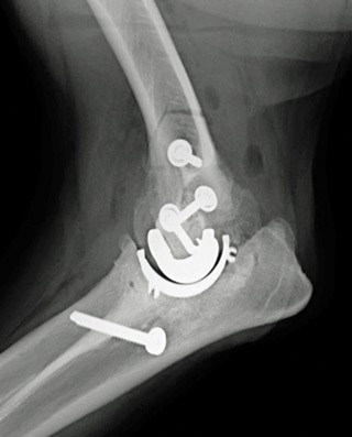
The elbow joint is a much more complex joint than the hip. This has resulted in much slower implant development, as well as less predictable outcomes following surgery than that associated with THR (Conzemius et al, 2001; Conzemius, 2009; Acker 2009; Innes et al, 2012).
An increasing amount of evidence (both anecdotal and in the literature) suggests elbow replacement surgery offers benefits in carefully selected cases of elbow pain associated with OA.
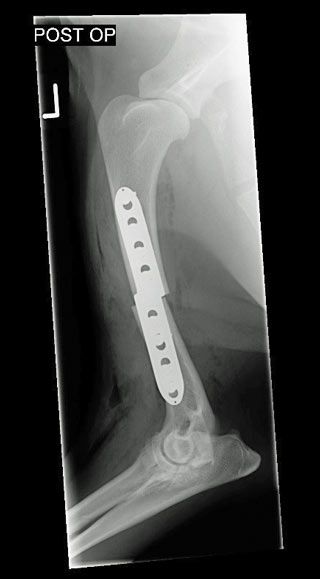
In the majority of cases it is preferable to arthrodesis. Objective data, including large numbers of cases of the elbow replacement systems available, is unfortunately lacking (Figure 2).
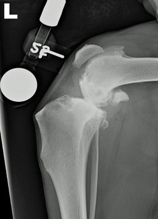
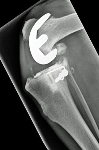
Total knee replacement (TKR) surgery for canine patients is another relatively recent innovation.
Reported case numbers are low (Liska et al, 2007; Eskelinen et al, 2012; Burton et al, 2014); however, an increasing number of UK referral centres offer this surgery and there is likely to continue to be an increasing amount of evidence to support TKR use in appropriate cases of, and advanced, stifle OA (Figure 3).
Excision arthroplasty
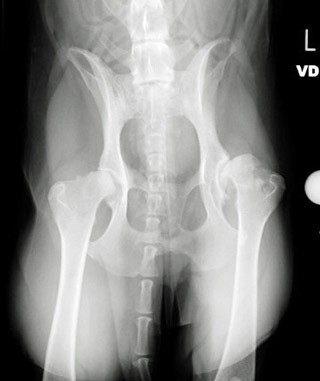
Excision arthroplasty of the coxofemoral joint (femoral head and neck excision; FHNE) is a commonly performed procedure for the treatment of painful hip joint conditions. When performed appropriately, FHNE results in reduced joint pain associated with OA.
Off and Matis (1997) reported functional results as good in 38% of patients, satisfactory in 20% and poor in 42% of cases. The outcome is not as reliable or predictable as that of a THR in dogs of any size and is therefore not the preferred treatment option for hip OA. It is, however, a cheaper and simpler procedure.
With good surgical technique (removal of the entire femoral neck) and postoperative analgesia and physiotherapy many small dogs do obtain reasonable limb function. The main complications of FHNE include ongoing lameness, limb shortening, muscle atrophy and decreased range of movements. Traditionally, it has been reported cats and small dogs can do very well following this surgery, but reports demonstrate THR is also a superior technique in these patients (Liska, 2010; Figure 4).
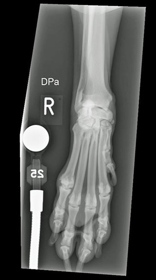
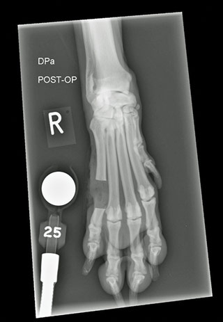
In the authors’ experience, excision arthroplasty can be a useful technique for the treatment of metacarpal/metatarsal joint or interphalangeal joint OA. It is a simple and quick cosmetically aesthetic alternative to arthrodesis (which, in the authors’ experience, is a challenging technique) or amputation of these joints (Jauernig et al, 1999).
The authors have seen a number of cases with persistent lameness localised to the metatarsal phalangeal joints respond favourably to this surgery (Figure 5).
Excision arthroplasty has also been reported as an effective alternative salvage procedure to arthrodesis for conditions of the canine shoulder joint (Bruecker and Piermattei, 1988).
Arthrodesis
The aim of joint arthrodesis is to achieve complete fusion of a joint at an appropriate standing angle. Fusion removes the discomfort associated with movement and can, therefore, be an effective treatment for dogs affected with painful OA.
The procedure is most commonly considered for management of pain associated with the carpi and tarsi. It can also be considered, although less commonly used, for intractable pain associated with the elbow, stifle and metacarpophalangeal and metatarsophalangeal joints.
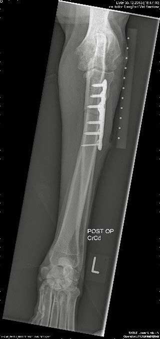
Pancarpal arthrodesis (rather than partial carpal arthrodesis) is most commonly indicated for OA of the carpal joint as the radiocarpal joint is typically involved. Limb function following surgery is generally good (Jerram et al, 2009; Sawyere et al, 2015; Bristow et al, 2015).
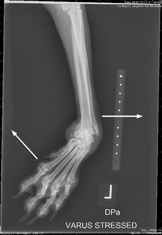
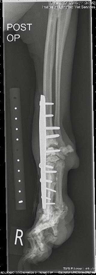
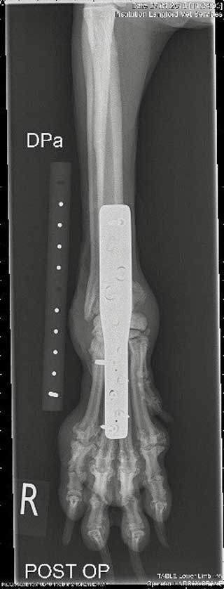
Advanced OA is a common problem affecting the hock joint secondary and commonly occurs as a result of osteochondrosis. Where the tibiotarsal joint is involved, pantarsal arthrodesis has been reported to give an acceptable outcome to most owners (McKee et al, 2004). Where the tibiotarsal joint is not involved, a partial tarsal arthrodesis is the preferred option.
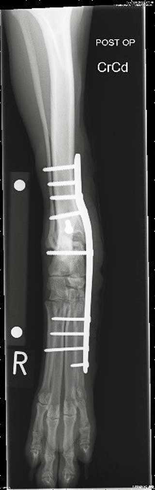
Partial tarsal arthrodesis typically results in excellent limb function as the motion of the talocrural joint is preserved (Figure 7).
Shoulder arthrodesis is an option for the management of shoulder OA poorly responsive to conservative treatment. The procedure is reported to result in surprisingly little functional impairment in dogs. This is most likely a result of a compensatory increase in the amount of scapula motion that occurs during locomotion following this procedure.
Pucheu and Duhautois (2008) reported shoulder arthrodesis results in good to excellent functional results in dogs in 87.5% of cases. In the authors’ experience, many cases of shoulder arthritis respond favourably to conservative therapy and, therefore, surgery has not been necessary or indicated.
Elbow arthrodesis is a salvage procedure for dogs with severe OA (Piermattei et al, 2006), but is typically reserved for cases where elbow replacement is not an option or has failed. Anecdotally, the procedure can result in relatively satisfactory function (Figure 8).
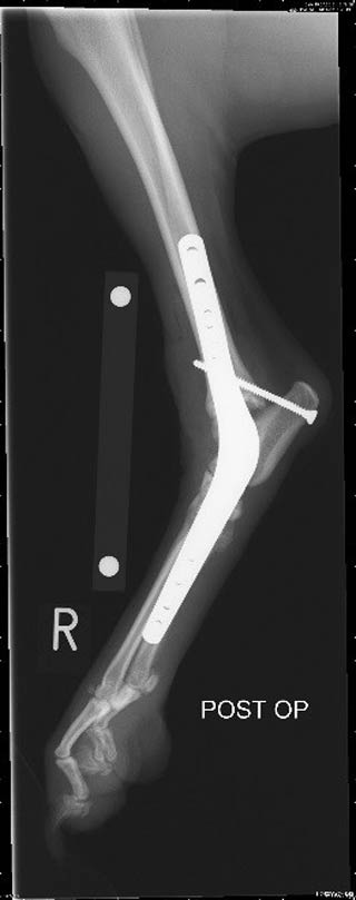
Stifle arthrodesis is an alternative to TKR where the latter is not an option (for example, if a dog is too small for a knee replacement). Reported limb function in these cases can be satisfactory; however, knee replacement is a preferable option whenever possible.
Arthroscopy and osteotomy procedures
Arthroscopy can be used as both a diagnostic and therapeutic tool. Arthroscopic debridement of bone fragments from within osteoarthritic joints can result in clinical improvement. This may be maintained in the long-term and the ongoing effectiveness will be dependent on the progression of arthritis within the joint.
A number of osteotomy techniques have been developed and applied to the elbow joint, including the proximal ulna osteotomy, sliding humeral osteotomy (Fitzpatrick et al, 2009; Wendelburg and Beale; 2014) and proximal abducting ulna osteotomy (Kyon, Switzerland). These procedures are often combined with arthroscopic assessment and/or treatment.
Elbow osteotomy procedures are aimed at altering the distribution of load across the articular surface and may improve function and comfort in patients where cartilaginous erosion is localised to the medial compartment of the elbow joint.
The long-term efficacy of these procedures is variable and the effect on future development of OA is as yet largely undetermined (Figure 9).
Conclusions
An increasing array of surgical options is available for the management of patients with OA. There are advantages and disadvantages to surgical options available.
A full discussion should be held with pet owners early in the course of the condition so they are fully informed of the choices available for ongoing treatment of their pet and, in some cases, early surgery may be beneficial.
Each case should be considered individually based on the pet’s response to previous treatments, owners’ expectations, patient factors and the joint involved. In patients proving challenging to manage, referral to an orthopaedic or pain clinic for specialist advice about surgical options and/or pain management is advised.
Acknowledgements
The authors would like to thank Peter Attenburrow from St David’s Veterinary Referrals in Exeter, for supplying the radiographs for this article.
Latest news
