19 Oct 2015
Radiotherapy in practice – part 2: uses and outcomes

Figure 3b. An MRI of the dog’s head showing a large pituitary tumour. This dog was treated with radiation therapy.
In the first part of this article, the principles of radiation biology, treatment planning and delivery were discussed. Part two offers an insight into the clinical application of radiation therapy in veterinary medicine, with a focus on the most commonly treated canine and feline neoplasms.
In small animal practice, radiation therapy plays an increasingly more important role in cancer treatment. Examples follow of canine and feline tumours commonly seen in veterinary practice that may benefit from radiation therapy.
Intranasal tumours
Radiation therapy is very important in the management of nasal tumours, which account for about 1% of all canine tumours. The majority (80%) are malignant (epithelial, mesenchymal, or round cell tumours) and carry a poor long-term prognosis1.


Even though the incidence of these tumours in animal patients is low, they are the fifth most common neoplasm treated with external beam radiotherapy2. The main problem with intranasal tumours is they are locally invasive and difficult to excise surgically although the risk of distant metastasis is minimal. If not treated, euthanasia is advised within a few months after the diagnosis due to progression of local disease and inability to control the associated clinical signs, particularly sneezing, nasal congestion and epistaxis3.
Radiation is the treatment of choice, with reported survival times for conventional radiotherapy ranging between 195 and 591 days4-6. More advanced radiotherapy techniques could potentially be used in the treatment of canine nasal tumours, such as intensity modulated radiation therapy or proton therapy7,8, and the outcome may be improved with these techniques; however, little data exists on the survival of these patients. Therefore, conventional conformal radiotherapy treatment is the most common treatment in canine nasal tumours – this may be prescribed as a definitive or palliative treatment.
The first approach is associated with moderate to severe long-term side effects, including keratitis, which, because cure in these dogs cannot be achieved and the long-term prognosis is poor, is not acceptable for the majority of owners.
In contrast, a study on 16 dogs with various nasal tumours revealed the dogs treated with definitive radiation (16-18×3Gy daily fractions to the total dose of 48Gy to 54Gy), followed by tumour exenteration, had no long-term side effects, and the median survival time of 457 days9. The main goal of a palliative approach is to alleviate the patient’s clinical signs (improve the airflow and stop epistaxis). Reports in the literature about use of radiotherapy in canine patients with nasal tumours differ in the fractionation schedule, dose per fraction and the total dose delivered. The median survival times ranged between 7 and 11 months10-12.
In a study evaluating 65 cats with various nasal tumours treated with radiotherapy, clinical signs improved in 86.2% of cats following radiotherapy. Acute complications, such as increase in sneezing, nasal and/or ocular discharge, were observed in more than half of the irradiated cats (58.5%), but were manageable and acceptable. Cataract was most frequently observed (20.5%) as the most common late side effect. The median overall survival and progression-free interval were 432 days and 229 days, respectively. There was no significant difference between overall survival for cats with nasal lymphoma compared with cats with other tumours13 (Figures 1a and 1b).

Intracranial tumours
Brain tumours are particularly frustrating and difficult to treat due to their location, lack of histopathological diagnosis in the vast majority of cases and tumours often being benign, slowly expanding lesions causing local compression of the healthy brain tissue. Complete surgical excision is almost always (with an exception of canine and feline forebrain meningiomas) impossible without causing an extensive damage to surrounding healthy brain tissue14. Palliative treatment with prednisolone results in a poor outcome15. Fractionated radiotherapy is a very useful treatment modality, either alone or as an adjuvant to surgery16-18. Various studies investigating various hypofractionated radiotherapy protocols for canine brain tumours have reported median survival times ranging from around 300 to 700 days17,18 (Figure 2).
Pituitary tumours
Both canine and feline pituitary tumours have also been treated with radiation therapy and, until recently in veterinary medicine, this constituted the treatment of choice in this type of tumour due to a different anatomy of the pituitary gland and more difficult and complicated surgical access compared to humans. A number of studies using different fractionation regimes for the treatment of pituitary macrotumours have reported in dogs. The survival times achieved with these protocols range from 147 days to more than 1,400 days18-23 (Table 1).

Radiation therapy has been reported to be an effective treatment for feline pituitary tumours, resulting in prolonged survival and control of both the tumour and paraneoplastic signs. In a study, five cats with pituitary tumours (four adenoma and one carcinoma) were treated with radiotherapy to a total dose of 39Gy prescribed in 12 fractions of 3.5Gy to 4Gy per fraction, delivered on a Monday-Wednesday-Friday schedule. The survival times were 5.5, 8, 15, 18 and 25 months24.
In a study on 12 cats with pituitary tumours treated with five weekly fractions to a total dose of 37Gy, the overall median survival time was 72.6 weeks25 (Figure 3a and 3b).


Soft tissue sarcoma
Soft tissue sarcomas originate from mesenchymal cells and represent approximately 15% to 20% of all cutaneous and subcutaneous tumours in the dog26.
Soft tissue sarcomas can arise in any anatomic site and most are contained within a “pseudocapsule”, which consists of compressed tumour cells surrounded by histologically poorly defined margins27. Their metastatic rate is low to moderate (15% to 41%), depending on the tumour grade, but locally aggressive behaviour is typical of soft tissue sarcomas28. Therefore, local tumour control is the most appropriate and important consideration in their management29.
The treatment of choice is complete surgical excision with surgical margin of 2cm to 3cm laterally and one deep fascia plane; however, as soft tissue sarcomas very often arise in anatomical areas with limited skin and soft tissue availability (distal limbs), complete excision is often not achieved, and additional therapy such as radiotherapy is indicated30.
Radiotherapy treatment may be advised pre or postoperatively; this very much depends on patient factors and available facilities. Neo-adjuvant radiation therapy has benefits (shrinkage of the tumour prior to surgery, less irradiated healthy tissue and so on); however, it is not commonly performed due to lack of sensitivity of macroscopic soft tissue sarcomas, most probably as a result of tumour hypoxia31.
Adjuvant radiotherapy for incompletely resected soft tissue sarcomas can be delivered in a form of definitive or palliative treatment. In one study, 35 dogs with soft tissue sarcoma were treated with a definitive protocol using 3Gy to 4.2Gy daily dose per fraction on a Monday to Friday schedule to a total dose of 42Gy to 57Gy. Overall median survival was 1,851 days and median time to recurrence was greater than 798 days32.
In another study on 48 dogs with soft tissue sarcomas treated with a Monday-Wednesday-Friday schedule, 3Gy/fraction/21 fractions to the total dose of 63Gy, five-year survival rate was 76%, median disease-free interval for all dogs was 1,082 days and median time to recurrence was 700 days30.
In contrast to these, another study on 56 dogs with soft tissue sarcoma treated postoperatively with hypofractionated radiotherapy protocol with a once-a-week schedule for four weeks, 8Gy to 9Gy/fraction to the total dose of 32Gy to 36Gy, documented a one-year, two-year, three-year and five-year-disease-free intervals of 82%, 74%, 70% and 65% respectively. This study demonstrates hypofractionated radiotherapy following intentional marginal excision of soft tissue sarcomas results in a good long-term clinical outcome, with low recurrence rates33 (Figures 4a to 4d).
It is important to mention marginally excised low-grade soft tissue sarcoma may do very well without any additional therapy34. Radiotherapy in an adjuvant setting for the treatment of previously excised soft tissue sarcoma needs to be part of a carefully formulated plan of treatment for the patient and not prescribed as an afterthought to salvage an inadequate surgery.
Canine mast cell tumours
Mast cell tumours (MCT) are another example of a very common canine neoplasm that presents a therapeutic challenge and can benefit from radiation therapy. MCT is the most common cutaneous tumour in the dog and it accounts for approximately 21% of all canine cutaneous tumours35. Similarly to soft tissue sarcomas, surgical excision is the treatment of choice for most cutaneous MCT, but locally recurrent, diffuse, or non-resectable MCTs represent a therapeutic challenge and warrant additional treatment.
Radiotherapy may be prescribed in the neo-adjuvant settings because the tumour is too large to be excised surgically, or is located in a very unfavourable place.
One of the possible complications of irradiating MCTs in situ, and a reason why this neo-adjuvant radiotherapy of MCTs is not commonly performed, is precipitation of histamine release by neoplastic mast cells. Use of prednisolone at high doses prior to radiation therapy is indicated to reduce the risk of degranulation and subsequent complications, and it may also facilitate surgical resection of many diffuse MCTs.
In a study on 35 dogs with non-resectable grade one to three MCT on the head or limbs, the patients were treated with 40mg/m2 prednisolone daily for 10 to 14 days prior to hypofractionated radiation therapy, which was delivered on a weekly schedule, 8Gy/fraction to the total dose of 32Gy. The overall response rate was 88.5%; the median progression-free interval was 1,031 days with one and two-year progression-free rates of 60% and 52%, respectively. Location, but not the grade of the tumour, influenced survival – dogs with tumours on the limbs survived longer than did dogs with tumours on their head36.
Hypofractionated radiotherapy in combination with toceranib and prednisolone has been prospectively evaluated in 17 dogs with non-resectable MCTs (12 with cytological diagnosis of MCT, five with histopathologically confirmed MCT, one dog with grade three, one dog with grade one MCT) or tumours not amenable to surgical excision. The dogs were treated with either four, once a week fractions of 6Gy/fraction to a total dose of 24Gy, or three once-a-week fractions of 8Gy/fraction to the same total dose – 24Gy. The overall response rate was 76.4%, with 58.8% of dogs achieving complete response and 17.6% a partial response; the median progression-free interval was 316 days, the median survival time was not reached and there was a median follow-up of 374 days37.
However, radiotherapy is most often implemented in adjuvant settings, once the tumour has been excised or cytoreductive surgery has been performed.


Many studies document good long-term outcomes for dogs with MCT treated postoperatively with radiotherapy. For example, in a study on 19 dogs with cutaneous MCTs and regional lymph node metastasis treated with cytoreductive surgery, prior to definitive radiotherapy consisting of a Monday to Friday schedule with 3Gy/fraction to the total dose of 48Gy to 57Gy, the median disease-free survival was 1,240 days38 (Figures 5a and 5b).
For more detailed medical treatment of canine MCT the reader is referred to an article from In Practice journal39.
Oral cavity tumours
Oral tumours present another therapeutic challenge due to their location and often advanced stage of disease at the time of presentation. The most common neoplasms in the oral cavity of a dog are oral malignant melanoma (OMM), fibrosarcoma, squamous cell carcinoma (SCC) and acanthomatous ameloblastoma. In cats, SCC predominates.
Radiotherapy can play an important role in an adjuvant setting in the management of oral tumours. Canine OMM is an extremely aggressive neoplasm with high degree of local invasiveness and high metastatic potential40. Loco-regional control is achieved by surgery and/or adjunctive radiotherapy, but even with control of the local disease recurrence rate is high: 70% of dogs will have a recurrent disease three to four months after surgical excision, and metastatic propensity is high also: 59% to 74% in regional lymph node, with 60% to 65% in lungs41.
Performing more radical surgical excision is therefore ethically questionable. This means radiotherapy may be more appealing to some clinicians and owners, and the more appropriate treatment option for these animals (including geriatric patients, concurrent diseases, high histological grade of the tumour).
Cutaneous and mucosal melanoma in humans responds better to a higher dose per fraction radiation schedule when compared to conventionally fractionated protocols; however, a consensus on the most appropriate radiotherapy dosing and scheduling does not exist in people42.
Hypofractionated radiation protocols have been used in primary treatment of OMM in veterinary medicine. In several studies response rates of greater than 80% have been observed with doses 8Gy to 9Gy per fraction. However, the overall median survival time ranged only between 5 to 7.9 months, which was due to metastatic disease43,44.
In a retrospective study on 140 dogs with OMM, dogs were treated with one of the three radiotherapy protocols. The median time to first event for all dogs was five months and the median survival time was seven months. There were no differences in response, time to first event and survival between the three radiation protocols45.
Oral fibrosarcoma is another common oral neoplasm; it is a mesenchymal tumour, composed of spindle cells or fibroblasts46. The treatment of choice, providing the best local control, is surgical excision followed by adjuvant radiotherapy. In a retrospective analysis of 65 dogs with oral fibrosarcoma, a combination of surgery and adjuvant radiotherapy resulted in a median survival time of 505 days, and progression-free survival of 301 days47.
Acanthomatous ameloblastomas (previously known as acanthomatous epulides) are benign oral tumours of odontogenic origin. They commonly invade the underlying bone so the surgical excision needs to be wide, including surrounding soft tissues and the underlying bone48. Complete surgical excision is curative. However, in some instances surgery is not possible (location or size of the tumour), or microscopic disease may remain following surgical resection, and that is where radiotherapy is indicated.
In a prospective clinical trial of 47 dogs with acanthomatous epulides, the dogs were treated with 12, 4Gy/fractions three times a week to a total dose of 48Gy. The estimate of three-year progression-free survival for these dogs was 80%49.

which was treated with radiation therapy.
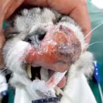
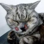
Squamous cell carcinoma is the most common oral tumour in cats. These tumours are locally invasive and most cats die early as a result of local disease50. Due to local extension of the tumour, surgery alone is not curative and response to radiotherapy is usually poor with relatively short survival times. Various retrospective studies describe different treatment protocols, but the number of cats in each study is small. Equally, the protocols prescribed (6Gy/fraction twice weekly; 8Gy/fraction on days 0, 7, 21; 4.2gy/fraction given on Monday-Wednesday-Friday schedule; 3.5Gy/fraction twice daily) and their application in either adjuvant (to surgery) or monotherapy setting, resulted in various survival times of 42.5 days, 116 days, 163 days, 174 days and 14 months51-56 (Figure 6).
Other tumours
Radiotherapy has been shown to be of palliative value in the management of histiocytic sarcoma (HS) and osteosarcoma.
Canine HS is an aggressive round cell tumour arising from malignant transformation of dendritic cells or macrophages57,58. Two forms – localised and disseminated – have been described. The localised form is most common in or around joints, skin, subcutaneous tissue and bone. This form is amenable to local therapies, but more than 90% of these dogs will develop distant metastases.
Radiotherapy may be useful as palliative management of this neoplasm to alleviate signs such as lameness, related to the primary mass. In a study of 37 dogs with HS, radiation therapy significantly improved survival. The median survival time of dogs treated with radiotherapy was 182 days versus 60 days for dogs that were not treated with radiation. A combination of CCNU and radiotherapy resulted in a median survival time of 208 days compared to 68 days for untreated dogs59.
Osteosarcoma is the most common primary bone tumour in dogs (85% of all primary bone tumours) and around 75% of such tumours affect the appendicular skeleton60. The prognosis is poor due to the aggressive biological behaviour and high metastatic potential of this tumour. The treatment of choice is limb amputation with adjuvant carboplatin chemotherapy.


Radiotherapy is an effective modality for temporary control of pain associated with primary and metastatic bony neoplasia in humans and dogs. Various approaches to palliative radiotherapy for the treatment of canine osteosarcoma have been described, with overall response rate ranging between 74% and 92%, median duration of response intervals ranging from 73 to 130 days, and median survival ranging from 122 to 313 days61,62. In most studies reported, clinical improvement is noticeable in approximately 50% of dogs by day 1462,63 (Figures 7a and 7b).
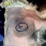
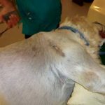
Feline injection site sarcomas (FISS, also referred to as vaccine-associated sarcomas) are aggressive, high-grade tumours thought to arise at injection sites and, due to their locally infiltrative growth, cause considerable difficulties in clinical management. Achieving long-term local control is difficult.
The most effective therapeutic approach is by a combination of surgical resection with radiation therapy. However, even when the tumour is fully excised with complete histological margins and full course, fractionated radiation therapy is applied, local tumour control is disappointing, with 28% to 45% recurrence rates following combination of these treatments64,65.
In a study on 92 cats with ISS, preoperative radiation therapy with 16 daily fractions of 3Gy/fraction to a total dose of 48Gy, median time to first event, which is the time calculated from the first day of treatment to local recurrence or metastatic disease, for all cats was 584 days, with only completeness of surgical excision being related to the time to first event. Median time to first event in the cats having had complete surgical excision was 986 days, compared to 292 days in cats with incomplete excision64.
This study demonstrated preoperative irradiation is an effective treatment for cats with ISS, especially if complete excision can be achieved following radiation therapy. It is incredibly important to appropriately plan the therapeutic approach in these cats before beginning any treatment.
Conclusion
In conclusion, radiotherapy plays a very important role in the management of various canine and feline tumours. It is generally well tolerated with minimal to moderate side effects in normal tissues, which is dependent on the fractionation regime chosen. With the new radiation delivery techniques, planning systems and equipment, these side effects can be reduced to a minimum.
Radiotherapy should be considered as a neo or adjuvant treatment in appropriately selected and suitable animal oncology patients. However, it is important to note when radiation therapy is used as adjuvant to surgery it needs to be part of a well-planned and thought through process and not an “additional” treatment to a poorly conceived surgical approach, which increases the likelihood and intensity of adverse effects and may result in treatment failure.
Latest news
