5 Nov 2024
Cytology findings from a skin nodule in adult dog
The latest in our Diagnostic Dilemmas series sees Francesco Cian delve into the case of an eight-year-old Maltese dog referred for the presence of a small cutaneous nodule on its neck.
Francesco Cian
Job Title
Figure 1. Skin mass in a dog (Wright-Giemsa 10× to 50×).
An eight-year-old Maltese dog was referred for the presence of a small cutaneous nodule on the back of the neck, which was noted during a grooming session.
The veterinarian performed a fine-needle aspirate (FNA) of the lesion and submitted the slides to an external laboratory for cytological examination.
The submitted smears are highly cellular with adequate preservation (Figure 1). The background is lightly basophilic with red blood cells. A main population of cuboidal epithelial cells arranged in clusters of variable sizes (black arrows) are visible. These cells have very small amounts of basophilic cytoplasm with poorly defined borders, small, hyperchromatic nuclei, and do not show signs of atypia.
Additionally, a few small individualised spindloid cells (red arrows) and small amounts of lightly pink amorphous material are visible. Very rare leukocytes are also noted.
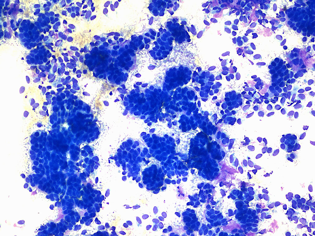
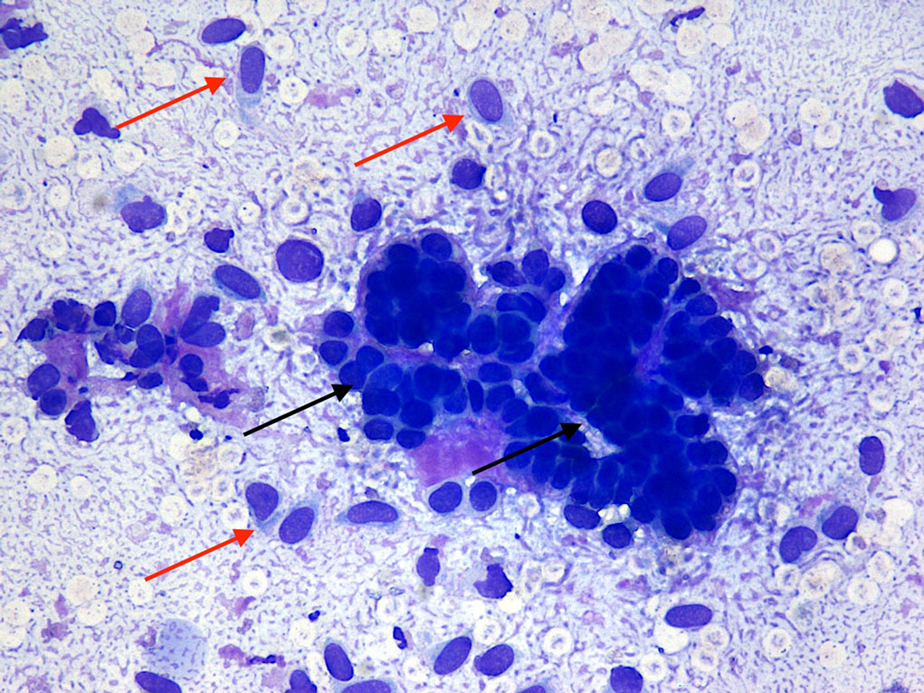
Figure 1. Skin mass in a dog (Wright-Giemsa 10× to 50×).
Diagnosis
A diagnosis of an adnexal epithelial tumour, likely a trichoblastoma, was found.
Insight
Adnexal tumours are a wide group of neoplasms arising from the adnexa, which include hair follicles, apocrine (sweat) and sebaceous glands.
The vast majority of these tumours have a benign clinical behaviour and exhibit cytological features that may partially overlap, hence the common use of this umbrella term.
Trichoblastoma, previously called a basal cell tumour, is one of the most common types of adnexal tumours and arises from the cells of the hair germ. It is particularly common in adult dogs, with poodles and setters likely being at increased risk.
It most often presents as a solitary, firm and alopecic mass, polipoid or dome-shaped on variable locations – particularly the head, neck and base of the ears. It has a peculiar cytological appearance, as described.
In the feline species, neoplastic cells of trichoblastoma may, at times, appear spindle, making differentiation from mesenchymal proliferation difficult. They may also contain melanin pigment, resulting in a pigmented nodule, making it challenging to clinically differentiate from a melanocytic tumour.
Trichoblastoma is a benign neoplasia and carries a good prognosis, with surgical excision considered to be curative.