31 Jul 2017
Dermatology hot spots in dogs – treatment and advice for owners
Jon Hardy
Job Title
Figure 1. Flea infestation and flea allergy dermatitis are common triggers for pyotraumatic dermatitis. This image shows abundant flea faeces from a coat brushing.
Pyotraumatic dermatitis, also called acute moist dermatitis or “hot spot”, is an acute onset, self-induced and traumatic skin disease caused by pruritus or pain. It is common in dogs, but rare in cats.
Despite its prevalence in dogs, very few published studies on the condition exist and many treatment recommendations are based on anecdotal experience.
Causes
Underlying causes are varied, but include all the differential diagnoses for pruritus in dogs. Fleas and flea allergy dermatitis are reported to be common triggers in dogs (Figure 1). Table 1 lists the most common triggers.
| Table 1. The most common triggers for pyotraumatic dermatitis (hot spots) in dogs | |
|---|---|
| Cause | Examples |
| Parasites | Fleas, lice, Sarcoptes scabiei, Otodectes cynotis, Neotrombicula autumnalis |
| Infections | Staphylococcal folliculitis, Malassezia dermatitis |
| Allergies | Flea allergy dermatitis, cutaneous adverse food reaction, atopic dermatitis |
| Otitis externa | |
| Anal sac disease | Infection, impaction, abscess |
| Foreign bodies in coat | Plant material |
| Irritant substances | |
| Poor/unkempt coat | |
| Underlying musculoskeletal disorders | Joint pain, soft tissue surgery |
Clinical signs
Pyotraumatic dermatitis has a rapid onset, with signs usually being noticed by owners within 72 hours (Holm et al, 2004). A typical lesion develops due to licking, chewing and scratching, and consists of well demarcated erythema, exudation and alopecia.
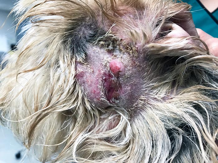
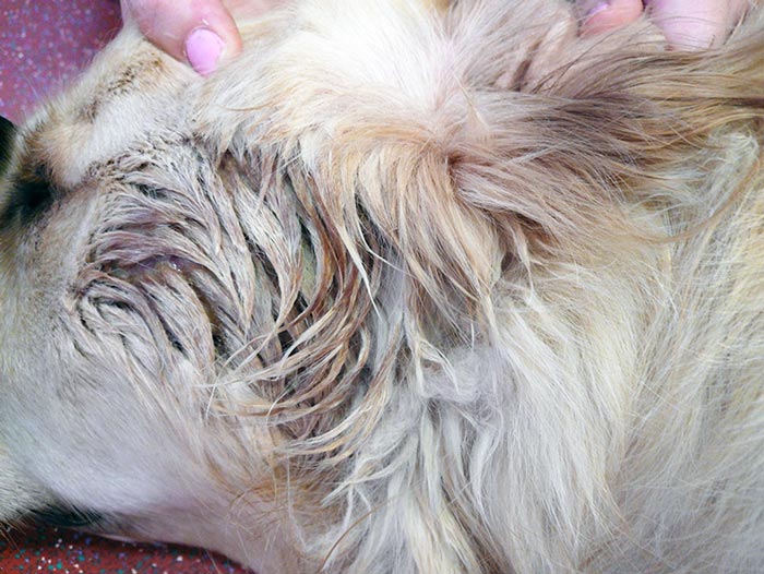
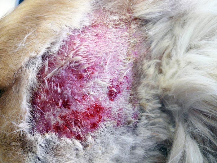
With time, erosions and ulceration form and proteinaceous debris accumulates on the skin surface and mats the surrounding hair (Figures 2 to 4). Lesions are often solitary, but can be multiple, and rapid progression occurs unless treatment is started. Pain and/or pruritus are consistent features.
Pyotraumatic dermatitis lesions often have surface bacterial colonisation due to Staphylococcus pseudintermedius (Holm et al, 2004), but are not true skin infections.
Pyotraumatic dermatitis therefore differs from pyotraumatic folliculitis and furunculosis, which involves bacterial infection of hair follicles and is classified as a pyoderma. The latter typically causes thickening and more plaque-like lesions.
In addition, satellite lesions of papules and pustules are often seen, which contrasts the well demarcated lesions of pyotraumatic dermatitis. Despite this, a study assessing the histopathological features of both diseases concluded it was difficult to differentiate the two, based purely on clinical signs (Holm et al, 2004).
Where does pyotraumatic dermatitis develop?
Pyotraumatic dermatitis usually forms near the site of the primary trigger. As flea allergy dermatitis is a common underlying cause, lesions are often found over the dorsal lumbosacral region and lateral thighs. The rump and skin around the anus are common sites when the anal sacs are involved and the skin around the ears is often affected when otitis is triggering the problem.
The lateral neck near the ears is also a common location for pyotraumatic folliculitis and furunculosis. When allergic skin disease, such as atopic dermatitis, is the trigger, lesions can be in a variety of locations.
Predisposing factors
Pyotraumatic dermatitis tends to be seen more commonly in large-breed dogs with dense coats. Breeds such as the German shepherd dog, golden retriever, Labrador retriever, collie, St Bernard and Rottweiler are commonly affected, although all dogs can create the lesion (Holm et al, 2004; Miller et al, 2013).
One study found dogs younger than four years of age and male dogs were more likely to be affected (Holm et al, 2004). However, other texts have reported no age or sex predilections. Pyotraumatic dermatitis may be more common during periods of warm and humid weather, but cases can occur throughout the year (Holm et al, 2004).
Diagnosis
Diagnosis is usually made based on the history ofacute onset disease, the distinctive clinical appearance, location of the lesion and the identification of a primary trigger.
Palpation of the skin is also useful and usually reveals a flat and well demarcated lesion.
Skin biopsy is not commonly required, but may be useful for recurrent or chronic cases and can help rule out differential diagnoses, such as ulcerated neoplastic lesions and pyotraumatic folliculitis and furunculosis.
Histopathology
Most changes are limited to the epidermis and include erosion/ulceration and surface exudation with crusts. Surface colonisation of these crusts with staphylococci is common. Lesions are usually very well demarcated from the adjacent epidermis, which may be spongiotic (oedematous).
The dermis often shows mild neutrophilic and lymphohistiocytic inflammation (Gross et al, 2005; Holm et al, 2004; Reinke et al, 1987).
Treatmentand prevention
Treatment needs to be given promptly to prevent expansion of the lesion and worsening of the pruritus/pain. Owners should, therefore, be advised to present their dog for a consultation as soon as possible. In addition, owners need to understand the lesion has formed due to a pruritic/painful trigger, and that identifying and correcting this is a vital part of management.
Initial treatment involves cleaning the lesion with a topical antimicrobial, such as chlorhexidine. Shampoo formulations are usually best, although sprays, wipes, gels and pads are available. Clipping hair aids cleaning, as well as removal of matted hair and proteinaceous debris from the lesion’s surface. In advanced cases, clipping and cleaning can be painful, so sedation may be required. This emphasises the importance of owners presenting pets for consultation at the earliest opportunity.
Following cleaning, the need for further treatment will depend on the individual case. Some cases benefit from anti-inflammatory doses of corticosteroids. Oral corticosteroids have the advantage that further manipulation of the lesion is not required, but the use of a systemic corticosteroid for a very focal problem is not ideal. For this reason, topical corticosteroids, such as a 0.584mg/ml hydrocortisone aceponate spray or fusidic acid/betamethasone gel, are often used (both available as licensed products in the UK).
The latter was shown in a study to be as effective as parenteral dexamethasone and oral clavulanate-potentiated amoxicillin after a seven-day period (Cobb et al, 2005).
An older study investigated the topical application of a neomycin-prednisolone combined product, and found the combination was more effective than when the component drugs were used alone (Schroeder et al, 1996). This product is not licensed for dogs in the UK.
Oclacitinib may also be useful in some cases – especially those with underlying allergic skin disease.
Some cases benefit from ongoing analgesia – especially if topical treatments are continued at home. NSAIDs can be useful for pyotraumatic dermatitis (Viking Höglund and Frendin, 2002).
Systemic antibiotics are usually not needed unless thickening of the skin and satellite lesions are present to suggest follicular involvement, and pyotraumatic folliculitis and furunculosis. If the patient is continuing to lick and bite at the area, Elizabethan collars may be required to limit further self-trauma.
When these collars are impractical or not tolerated, T-shirts or body suits may be used. In general, treatment for pyotraumatic dermatitis usually lasts for around 7 to 14 days.
Along with management of the lesion, which is normally fairly straightforward, a thorough search for the underlying trigger is required, which may involve several in-house diagnostic tests (Table 2).
| Table 2. In-house diagnostic tests that may be required when investigating cases of pyotraumatic dermatitis | |
|---|---|
| Diagnostic test | Disease |
| Coat brushing | Flea infestation, cheyletiellosis, pediculosis, trombiculosis, otoacariasis |
| Hair plucks | Demodicosis, dermatophytosis |
| Skin scrapings | Demodicosis, sarcoptic mange, cheyletiellosis |
| Tape impressions/impression smears | Malassezia dermatitis bacterial folliculitis |
| Cytology swabs of otic cerumen | Malassezia and bacterial infections of ears |
The ears and anal sacs may need to be evaluated and the condition of the coat should be assessed. A full dermatological examination should be performed to look for signs of allergic skin disease.
In some animals, imaging of the underlying joints/bones may be required if a painful focus is suspected.
Identification of the trigger is key to preventing future lesions, as prophylactic measures can then be put in place. This may involve regular ectoparasite control or regular emptying of anal sacs in dogs prone to anal sacculitis.
Owners may need to improve their grooming and clipping regimen to ensure the coat remains free of matts and foreign material. This may be even more important in hot and/or humid weather.
In cases where otitis has triggered pyotraumatic dermatitis, it may be necessary for owners to clean the ears regularly with a suitable general purpose ear cleaner.
If the patient has underlying allergic skin disease, control over this disease will be key to preventing future relapses.
Conclusion
Identification and treatment of pyotraumatic dermatitis is not normally problematic. However, the challenge is to identify the trigger and then put preventive measures in place.
The success of this relies on owners understanding the aetiopathogenesis of pyotraumatic dermatitis and the importance of preventing pruritic/painful triggers.
References
- Holm BR, Rest JR and Seewald W (2004). A prospective study of the clinical findings, treatment and histopathology of 44 cases of pyotraumatic dermatitis, Veterinary Dermatology 15(6): 369-376.
- Miller WH, Griffin CE and Campbell KL (2013). Environmental skin diseases. In Muller and Kirk’s Small Animal Dermatology (7th edn), Elsevier, St Louis: 677-678.
- Cobb MA, Edwards HJ, Jagger TD, Marshall J and Bowker KE (2005). Topical fusidic acid/betamethasone-containing gel compared to systemic therapy in the treatment of canine acute moist dermatitis, The Veterinary Journal 169(2): 276-280.
- Gross TL, Ihrke PJ, Walder EJ and Affolter VK (2005). Ulcerative and crusting diseases of the epidermis. In Skin Diseases of the Dog and Cat: Clinical and Histopathologic Diagnosis (2nd edn), Blackwell Science, Oxford: 116-118.
- Reinke SI, Stannard AA, Ihrke PJ and Reinke JD (1987). Histopathological features of pyotraumatic dermatitis, Journal of the American Veterinary Medical Association 190(1): 57-60.
- Viking Höglund O and Frendin J (2002). Analgesic effect of meloxicam in canine acute dermatitis – a pilot study, Acta Veterinaria Scandinavica 43(4): 247-252.
- Schroeder H, Swan GE, Berry WL and Pearson J (1996). Efficacy of a topical antimicrobial-anti-inflammatory combination in the treatment of pyotraumatic dermatitis in dogs, Veterinary Dermatology 7(3): 163-170.
Meet the authors
Jon Hardy
Job Title