17 Apr 2023
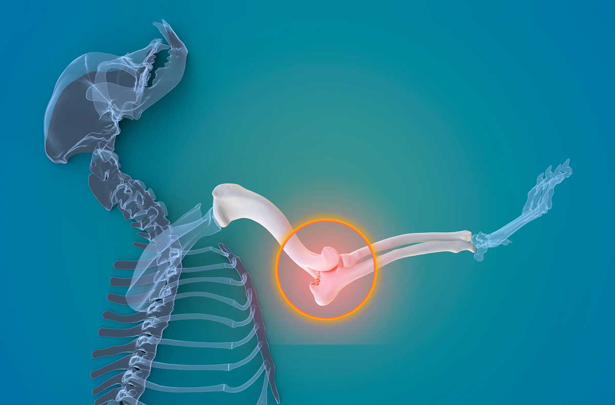
Elbow dysplasia – an update
Zoë Nalborczyk BSc, BVMSci, MRCVS and Rob Pettitt BVSc, PGCertLTHE DSAS(Orth), SFHEA, FRCVS discusses the latest treatment options for this orthopaedic condition in dogs.

Image: © 9gifts / Adobe Stock
Elbow dysplasia – more recently termed developmental elbow disease – is a collective phrase encompassing developmental abnormalities of the humeroradialulnar joint.
These are medial coronoid process disease (MCPD), osteochondritis dissecans of the medial humeral condyle (OCD), elbow joint incongruity and ununited anconeal process (UAP), which can occur in isolation or in association with each other, with joint incongruity contributing to the development of MCPD and UAP (Michelsen, 2013; Samoy et al, 2006).
Elbow dysplasia is most common in large and giant-breed dogs, including the Labrador retriever, German Shepherd dog, golden retriever, Rottweiler, Bernese mountain dog, Newfoundland, great Dane and dogue de Bordeaux (Kirberger and Fourie, 1998; LaFont et al, 2002), although smaller breeds such as cavalier King Charles spaniels are becoming more commonly affected.
Elbow dysplasia can significantly impact quality of life and is characterised by pain, lameness, reduced range of motion and reluctance to exercise.
Components of elbow dysplasia
Elbow joint incongruity
Elbow joint incongruity is a broad term that encapsulates misalignment of the humeroradial, humeroulnar and radioulnar joints, which together compose the elbow joint.
The two most common forms are humeroulnar incongruence and radioulnar incongruence. Humeroulnar incongruence results from the abnormal formation of the trochlear notch (Eljack and Bottcher, 2015).
Radioulnar incongruence occurs due to a mismatch in growth between the ulna and radius, resulting in either a short radius (known as positive incongruence) or a short ulna (known as negative incongruence), and results in humeroulnar conflict (Wind, 1986a, Wind, 1986b; Eljack and Bottcher, 2015).
The consequence of elbow joint incongruity is abnormal load distribution across the joint and subsequent overloading of the medial compartment, resulting in cartilage lesions and degenerative joint disease (Fitzpatrick and Yeadon, 2009; Fitzpatrick, 2013; Michelsen, 2013).
Elbow joint incongruity has also been associated with the pathogenesis of medial coronoid process disease and ununited anconeal process. The condition is most commonly seen in large to giant breed dogs including Labrador retrievers, German shepherd dogs, golden retrievers, Rottweilers and Bernese mountain dogs; however, smaller chondrodystrophic breeds are also predisposed (Lavrijsen et al, 2012; Eljack and Bottcher, 2015).
Medial coronoid process disease
Medial coronoid process disease is the most prevalent component of elbow dysplasia and associated with joint incongruity causing biomechanical overload of the medial coronoid process during maturation (Wind, 1986a; Wind 1986b; Samoy et al, 2006; Fitzpatrick et al, 2016).
This results in sclerosis and microfractures of the subchondral bone, which propagate, causing fissure formation and fragmentation of the medial coronoid process in addition to erosion of the articular cartilage.
Overt joint incongruity is not always noted at the time of diagnosis of MCPD, which is attributed to resolution of the incongruity during residual growth of the patient (Coppieters et al, 2015). MCPD is most common in Labrador retrievers, German shepherd dogs and Rottweilers, and is twice more prevalent in males (Fitzpatrick et al, 2009a).
Osteochondritis dissecans
Osteochondritis dissecans of the medial condyle occurs due to focal disruption of endochondral ossification causing abnormally thickened cartilage and resulting in ischaemic necrosis. This results in the formation of a fissure in the articular cartilage, which progresses to form an osteochondral flap and exposure of the subchondral bone, resulting in inflammation and degenerative joint disease.
OCD occurs most frequently in Labrador retrievers, golden retrievers, Bernese mountain dogs and Newfoundlands, and is more commonly reported in males (Bennett et al, 1981; LaFont et al, 2002; Vezzoni and Benjamino, 2021).
Ununited anconeal process
Timing of ossification of the anconeal process varies between breeds, with failure to ossify by 20 weeks of age classified as an ununited anconeal process (Van Sickle, 1966; Breit et al, 2004; Vezzoni and Benjamino, 2021).
The underlying pathophysiology of ununited anconeal process is unclear, with asynchronous growth of the radius and ulna, and resulting joint incongruity most widely accepted, causing abnormal pressures on the anconeal process (Vezzoni and Benjamino, 2021).
The functional implications of an ununited anconeal process are increased joint instability due to the presence of a fragment within the joint and loss of the stabilising function of the anconeal process, resulting in cartilage erosion and degenerative joint disease (Fox et al, 1996).
The condition occurs bilaterally in 20% to 35% of cases and males are twice as frequently affected (Hayes et al, 1979; Sjöström, 1998). Although historically considered to be a disease of large and giant breed dogs, ununited anconeal process has been reported in the French bulldog and dachshund (LaFont et al, 2002).
Medial compartment disease
Three of the four components of developmental elbow disease – MCPD, OCD and joint incongruity – affect the medial compartment of the elbow joint and are often concurrent.
These disease processes result in inflammation and erosion of the articular cartilage with subsequent progression of degenerative joint disease and OA, which is collectively termed medial compartment disease (Fitzpatrick, 2013; Bruecker et al, 2021).
Treatment of elbow dysplasia
Conservative management
The principles of conservative management of medial compartment disease are the use of non-surgical strategies to minimise lameness and discomfort, improve function and minimise the rate of progression of degenerative joint disease.
The components of conservative management of medial compartment disease are similar to that of conservative management of any osteoarthritic joint and consist of analgesia, weight management and exercise modification. Adjunctive physiotherapy and hydrotherapy are recommended to preserve muscle mass and range of motion of affected joints.
Use of joint supplements containing omega-3 fatty acids is supported within the literature; however, evidence regarding the benefits of other supplements are lacking (Roush et al, 2010; Fritsch et al, 2010).
Weight reduction has been associated with improvements in lameness and mobility in multiple studies (Impellizeri et al, 2000; Mlacnik et al, 2006; Marshall et al, 2010). This is attributed to reduced loading through osteoarthritic joints and reduced adipose-derived inflammatory mediators (Ouchi et al, 2011).
Patients with elbow dysplasia should be kept lean with an ideal body condition score of 4/9 and overweight patients should set an initial target of 15% bodyweight loss.
Communication with owners regarding the importance of weight management is crucial. Owners of overweight patients should be provided with guidance with regards to weight reduction strategies, with the use of nurse-led weight management clinics often extremely useful.
Exercise modification is a balance between preventing aggravation of osteoarthritic joints, while maintaining quality of life. High-intensity activities involving turning at speed and sharp breaking, such as ball chasing and boisterous play with other dogs, should be avoided.
Owners commonly have the misconception that a dog will not exercise if it is in pain; therefore, communicating to owners that patients are unable to self-regulate to associate increased activity with increased discomfort – and thus, the responsibility on owners to control exercise – is crucial.
During periods of acute exacerbations (“flare-ups”), exercise should be markedly reduced and controlled, alongside the use of NSAIDs to manage inflammation and minimise the duration of the flare-up. Due to the variations in severity of lameness and other patient factors, a standard exercise regime is difficult to recommend.
The principles of acute flare-up exercise management are frequent short walks controlled on the lead, alongside restriction within the home, preventing energetic behaviour in the garden and with other dogs. Jumping on and off furniture, and into and out of vehicles, should also be prevented, as well as climbing stairs.
As the flare-up resolves and the patient’s lameness improves, the length of on-lead walks can be gradually increased to their previous levels of exercise followed by the introduction of off-lead exercise towards the ends of walks.
NSAIDs can be used intermittently, or continuously to manage the inflammation and pain associated with acute exacerbations of lameness, and chronic pain from degenerative joint disease. Additional analgesia can be implemented in patients with limited response to NSAIDs, including paracetamol and amantadine.
Tramadol has poor evidence regarding efficacy in dogs, with only one of its eight metabolised substrates active in the dog for one to two hours (Delgado et al, 2014; Benitez et al, 2015).
Bedinvetmab has been introduced for the management of osteoarthritic pain; however, use of bedinvetmab in combination with NSAIDs is controversial as a lack of safety studies exist regarding concurrent use in dogs, and studies in humans have reported rapidly progressive OA associated with concurrent monoclonal antibody therapy and NSAIDs.
Limited reports exist of the use of intra-articular injections of a range of substances including corticosteroids, platelet-rich plasma and autologous stem cells (Fahie et al, 2013; Franklin and Cook, 2013; Wanstrath et al, 2016).
Further studies into their therapeutic effectiveness is warranted; however, their limited duration of action necessitates the requirement for repeated injections, which restricts their application due to the associated costs and requirement for repeat procedures under sedation.
Stem cells in particular are logistically more challenging and invasive, requiring harvesting from the patient’s adipose tissue – often from the falciform fat – requiring a midline coeliotomy under general anaesthesia to obtain.
Surgical treatment of specific pathologies
Elbow joint incongruity and medial coronoid process disease
Dynamic ulnar osteotomies have been suggested to improve elbow congruity and prevent worsening of medial coronoid process disease in skeletally immature dogs; however, evidence demonstrating this is lacking.
Distal dynamic ulnar ostectomy (DDUO) has been performed in four to six-month-old dogs with suspicion of medial coronoid process disease due to the presence of subtrochlear sclerosis on radiographs (Vezzoni, 2000; 2014).
This involves removal of 4mm to 5mm of the ulna 2cm to 3cm proximally to the distal ulna physis, and is thought to remove supraphysiological pressure on the medial coronoid process by facilitating sliding of the ulna proximally or distally depending on the type of joint incongruity present (Ness, 1998, Preston et al, 2001; Bottcher et al, 2013).
Unpublished data from Vezzoni and Benjamino (2021) comparing DDUO with conservative management suggests DDUO is effective at reducing the progression of OA in most cases; however, peer-reviewed studies regarding outcomes and comparison with other treatment options are lacking.
Bi-oblique dynamic proximal ulnar osteotomy (BODPUO) has been performed in more severely affected puppies and skeletally immature dogs up to 12 months of age with cartilage damage to the medial compartment on arthroscopic assessment. Proximal ulnar osteotomy relies on tension and compression forces acting on the proximal ulna segment to improve joint congruence, and lateralises the manus to unload the medial compartment.
A bi-oblique direction to the osteotomy reduces excessive caudal and varus tilting of the proximal ulna segment, and reduces the occurrence of major complications. A retrospective case series of 120 elbows reported no major complications and 12% minor complications with significantly decreased subjective lameness scores for all dogs at six weeks postoperatively (Caron and Fitzpatrick, 2016).
Bilateral simultaneous BODPUO is associated with a higher incidence of postoperative complications; therefore, staged procedures are recommended.
Arthroscopic removal of fragmented medial coronoid process is the traditional treatment of medial coronoid process disease. The procedure entails removal of the visibly diseased portions of the medial coronoid process using arthroscopic graspers or mosquito forceps followed by debridement of the exposed sclerotic subchondral bone.
Subtotal coronoid ostectomy is the removal of the majority of the tip of the medial coronoid process to remove microcracks that can be present in the visibly healthy areas of the medial coronoid process to prevent recurrent fragmentation and persistent discomfort (Fitzpatrick et al, 2009a; Fitzpatrick et al, 2009b).
A prospective study in 263 dogs identified 71.5% of dogs were sound on subjective lameness assessment 12 weeks following surgery, with 81.9% of dogs remaining sound in the medium term (Fitzpatrick et al, 2009b).
Care must be taken with regards to the aggressiveness of subtotal coronoid ostectomy performed, with acute end-stage medial compartment disease reported in six dogs due to humeroulnar conflict from significant reduction in the weight bearing surface of the medial coronoid process (Bräuer and Bottcher, 2015).
Ultimately treatment of medial coronoid process disease by fragment removal and/or subtotal coronoid ostectomy does not address the underlying causes of medial coronoid disease with progression of medial compartment disease seen in most cases (Barthelemy et al, 2014; Vezzoni and Benjamino, 2021).
Factors influencing the decision to perform arthroscopic elbow surgery include the presence and type of osseous pathology – for example, fissured or fragmented medial coronoid process, the degree of OA, the age of the patient and the preference of the surgeon (Fitzpatrick and Yeadon, 2009).
The approach used by the authors for medial coronoid process disease in dogs is conservative management in the first instance, with arthroscopy considered when response to conservative management is deemed unsatisfactory. Even at this stage, a minimal approach is taken with regards to fissure removal and debridement of the adjacent cartilage.
Osteochondritis dissecans
Traditional treatment of osteochondritis dissecans lesions of the medial humeral condyle is arthroscopic debridement and abrasion arthroplasty. This involves debridement of the OCD cartilage flap followed by curettage or foraging of the subchondral bone to promote migration of mesenchymal stem cells, angiogenesis and healing of the defect in the cartilage with fibrocartilage.
Some dogs have been shown to improve clinically following surgery; however, chronic lameness has been reported in many cases with progression of degenerative joint disease (Bennett et al, 1981; Bouck et al, 1995; Boudrieau et al, 1983).
Osteochondral autogenous transfer involves removal of the OCD cartilage flap followed by placement of an autogenous osteochondral transplant harvested from the lateral aspect of the femoral trochlea (Fitzpatrick et al, 2009c). This allows reconstruction of the articular surface of the medial humeral condyle with hyaline cartilage; however, the technique requires surgery on a distant joint for transplant harvest, which risks donor site morbidity (Bottcher et al, 2009; Fitzpatrick et al, 2009c).
Use of osteochondral synthetic implants has been developed using thermoplastic polycarbonate urethane plugs to overcome the limitations of autografts (Cook et al, 2014). Unpublished data on clinical follow-up of elbow joint SynACart implantation in combination with BODPUO revealed no complications, with good to excellent function in the majority of cases (Preston, 2016).
A lack of peer-reviewed studies exists comparing the three procedures, which makes drawing conclusions regarding the most effective treatment method challenging.
Ununited anconeal process
Surgical excision of the anconeal process through a caudolateral approach to the elbow was traditionally used to treat an ununited anconeal process.
Prognosis following this procedure was variable between studies, with one retrospective study reporting 90% of owners rating the outcome of surgery as good to excellent with regards to their dogs’ activity level (Roy et al, 1994).
Conversely, another study reported only 50% of dogs were sound following surgery, with a reduction in the normal range of movement in the elbow to a mean of 68% in flexion and 84% in extension (Sinibaldi and Arnoczky, 1975).
The benefits of this procedure are likely to be limited due to failure to resolve the underlying joint incongruity and instability – and, therefore, progression of degenerative joint disease (Roy et al, 1994; Vezzoni and Benjamino, 2021).
Accordingly uniting the fragments should be prioritised in skeletally immature dogs; however, surgical excision could be considered in skeletally mature dogs with degenerative joint disease.
Bi-oblique dynamic proximal ulna osteotomy is performed to manage the underlying joint incongruity thought to contribute to the development of an ununited anconeal process to facilitate union between the olecranon and the anconeal process (Sjöström et al, 1995).
Studies have demonstrated good to excellent clinical outcomes; however, the rate of union is variable, with a 71% union rate in one study and 21% union rate in another (Sjöström et al; 1995; Turner et al, 1998).
Reattachment of the anconeal process to the olecranon through placement of lag screws and/or Kirschner wires via a lateral approach to the elbow is an alternative treatment in dogs up to 24 weeks old with a normally formed trochlear notch. Precise positioning of the anconeal process is crucial to prevent implant failure through increased contact between the anconeal process and the humerus; however, underlying joint incongruity can make this challenging (Sjöström, 1998).
Accordingly, anconeal process reattachment can be performed concurrently with a bi-oblique dynamic proximal ulna osteotomy to restore the integrity, congruity and biomechanics of the elbow joint.
Several studies with a total of 58 elbows have demonstrated on overall fusion rate of 93% (Krotscheck et al, 2000; Meyer-Lindenberg et al, 2001). A further retrospective multicentre study comparing the outcomes of proximal ulna osteotomy alone and proximal ulna osteotomy with anconeal process reattachment found those elbows undergoing both procedures had a significantly better rates of radiographic union (84%; Pettitt et al, 2009).
Surgical management of medial compartment disease
For skeletally mature patients with mild to moderate medial compartment disease, two surgical procedures have been proposed to unload the medial compartment of the elbow joint to reduce joint pain from friction and prevent further progression of degenerative joint disease.
This is achieved through laterally shifting the weight-bearing axis of the thoracic limb to reduce load through the medial compartment of the elbow joint (Bruecker et al, 2021). These procedures are contraindicated in patients with pathology affecting the lateral compartment of the elbow and patients with severe degenerative joint disease (Bruecker et al, 2021).
Sliding humeral osteotomy (SHO) is a technique whereby a mid-diaphyseal humeral osteotomy is performed to translate the distal humeral fragment laterally and is secured with a custom locking plate on the medial aspect of the humeral diaphysis.
The procedure initially carried a high complication rate; however, further refinement of the custom implants resulted in a 19% overall complication rate, with 5% being major complications and improvement in lameness in 90% of cases (Wendelburg and Beale, 2014; Fitzpatrick et al, 2009d).
Introduction of a stepped plate to act as a guide for the osteotomy and sliding of the humerus was associated with no major complication and 4.17% minor complications in 60 cases (Fitzpatrick et al, 2015). Lameness was reported to be improved in all 60 cases by 12 weeks postoperatively and resolved in 49 cases. Growth of fibrocartilage in formerly eroded areas on the humeral trochlea has also been demonstrated following SHO, supporting unloading of the medial compartment (Wendelburg and Beale, 2014).
Proximal abducting ulnar osteotomy is a technique whereby a transverse proximal ulna osteotomy is performed and secured with a custom locking plate on the lateral aspect of the proximal ulna (Bruecker et al, 2021).
The plate has a step of 2mm to 3mm on the proximal aspect, which causes mild abduction, axial rotation and caudal tipping of the proximal segment of the ulna to shift the distal limb laterally (McConkey et al, 2016).
An unpublished case series of 116 cases reported a marked improvement in lameness (Vezzoni, 2018). Pre-operatively, 92% of cases were graded 2 to 3 out of 4 on a subjective lameness scale, with 8% graded 1 out of 4 and, of 49 cases assessed at 6 months postoperatively, 51% were graded sound and 40% were graded 1 out of 4.
A 4% major complication rate occurred, with plate removal required in all cases and a 2.5% minor complication rate (Vezzoni, 2018). A retrospective cohort study of 74 cases assessing postoperative complications was published last year and reported a 25% major complication rate, including non-union, implant failure and infection, with an increased likelihood of complications as bodyweight increased (Danielski et al, 2022).
For skeletally mature patients with end-stage medial compartment disease, characterised by full thickness cartilage loss with subsequent exposure of subchondral bone resulting in debilitating elbow pain from stimulation of subchondral bone nociceptors, joint resurfacing techniques have been developed.
The canine unicompartmental elbow (CUE) involves placement of a 4mm to 6mm polyethylene plug into the exposed medial coronoid process and a chrome prosthesis into the medial humeral trochlea. The principles of the CUE system are to limit bone on bone contact through provision of a low friction synthetic surface throughout the weight-bearing range of movement only, and to preserve the natural anatomic joint stabilisers to reduce the likelihood of instability or luxation postoperatively.
A prospective multicentre study of 103 cases using a medial approach to the elbow reported functional outcome to be full function in 47.6% of cases, acceptable function in 43.7% of cases and unacceptable function in 8.7% of cases, with 1 catastrophic, 11 major and 28 minor complications (Cook et al, 2015).
A retrospective case series of 52 cases using a caudomedial approach to the elbow reported functional outcome to be full function in 25% of cases, acceptable function in 73% of cases and unacceptable function in 1 case, with a 15% major complication rate consisting primarily of refractory pain and lameness (Bayer et al, 2019). The case series also reported the occurrence of implant-related contact lesions in eight cases when second-look arthroscopy was performed.
Total elbow replacement involves resurfacing of the entire elbow joint with prostheses. Two principal systems exist – Sirius, a semi-constrained hybrid system, and BioMedtrix TATE system, which is cementless. No published objective studies exist on the TATE system and limited long-term follow-up.
A retrospective multicentre case series of 33 elbows reported full functional outcome in 24% of cases and acceptable function in 52% of cases; however, 24% of cases had unacceptable function outcome (De Sousa et al, 2016).
A 15% rate of catastrophic complications occurred and a 45% occurrence of major complications, with suboptimal implant positioning in 97% of cases.
Decision making
The decision-making process for the treatment of elbow dysplasia remains controversial due to a lack of prospective clinical research with objective outcome measures for specific elbow dysplasia surgical procedures and treatments. Furthermore, few studies directly compare conservative management to surgical management of elbow dysplasia, and a lack of studies exists comparing different surgical treatments for elbow dysplasia.
Burton et al (2011) compared the outcomes of conservative management with dogs treated with arthroscopic fragment removal and found no difference in outcome one year later.
Given the relative paucity of evidence, the lack of consensus among existing evidence regarding the benefits of surgical management and the progression of degenerative joint disease despite surgical management, conservative management is recommended by the authors for treatment of elbow dysplasia in the first instance.
Surgical intervention can be considered if a patient’s response to conservative management is deemed insufficient; however, improvement following surgery is almost always temporary due to the inevitable progression of degenerative joint disease.
Take-home points
Take-home points include the following:
- The components of elbow dysplasia are closely inter-related in their pathogenesis and progression despite classification into four aspects.
- Non-surgical management is the mainstay of most cases consisting principally of weight management, exercise modification and NSAIDs.
- Arthroscopic removal of fragmented medial coronoid process can be considered in patients refractory to conservative management; however, postoperative improvement is inevitably temporary and care should be taken regarding the amount of medial coronoid removed.
- Surgical techniques to unload the medial compartment have been developed; however, a lack of studies exists regarding long-term outcomes with comparison of outcomes to surgical management.
- Joint resurfacing surgery can be considered in patients with advanced medial compartment disease; however, these techniques are still being developed and carry significant risks of complications with no effective revision options.
- Clear and effective communication with owners is crucial to set realistic expectations with regards to disease progression, degree of response to conservative management and the limitations of surgical interventions.
Latest news

Small animal
Evidence-based clinical nutrition to support weight management and joint care in dogs
Sponsored
28 Feb 2025