3 Nov 2022
Treatment of cataracts in a parrot
Richard Jones
Job Title
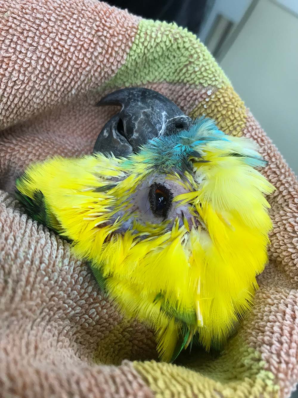
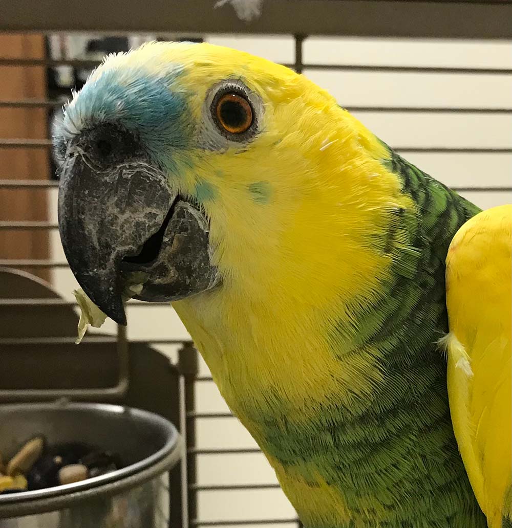
Figure 9. Ten weeks postoperatively with the patient now visual.
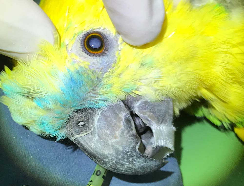
A juvenile blue-fronted Amazon parrot was presented to Avian Veterinary Services in Cheshire with assumed congenital cataracts that had unfortunately progressed to the point the bird was now blind and had, essentially, “shut down” – no longer able to negotiate or find food in its communal aviary (Figure 1).
Referral for specialist treatment was sadly not possible in this case on financial grounds and the author’s team did discuss euthanasia at this time, but elected to research other options that might be available to them in a general practice setting before going down that path.
They scoured the internet and came across the “sutureless fish hook technique”, developed for use in people “in the field” in remote areas of Nepal by Albrecht Hennig of the Lahan eye hospital. This technique essentially uses a hypodermic needle bent into a “fish hook”, then introduced via a scleral tunnel into the anterior chamber and used to hook out the lens nucleus. A similar technique had also been described in macaw species in Avian Medicine: Principles and Application (Ritchie et al, 1994).
Procedure
Following numerous cadaver trial runs and some tweaking of previous methods, the author’s team bit the bullet and went ahead with the surgery as follows under 3.5× magnification.
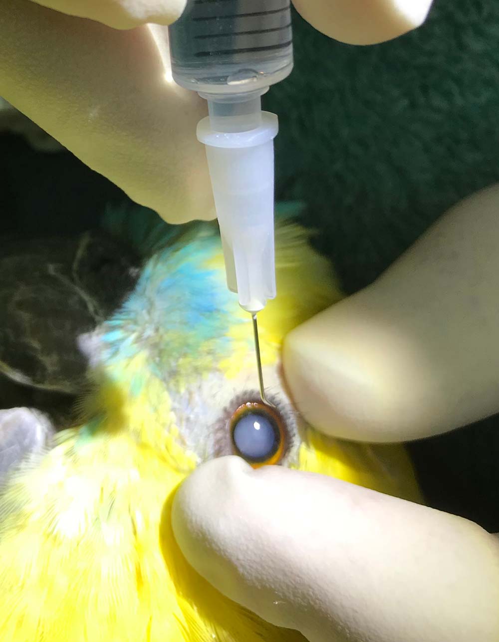
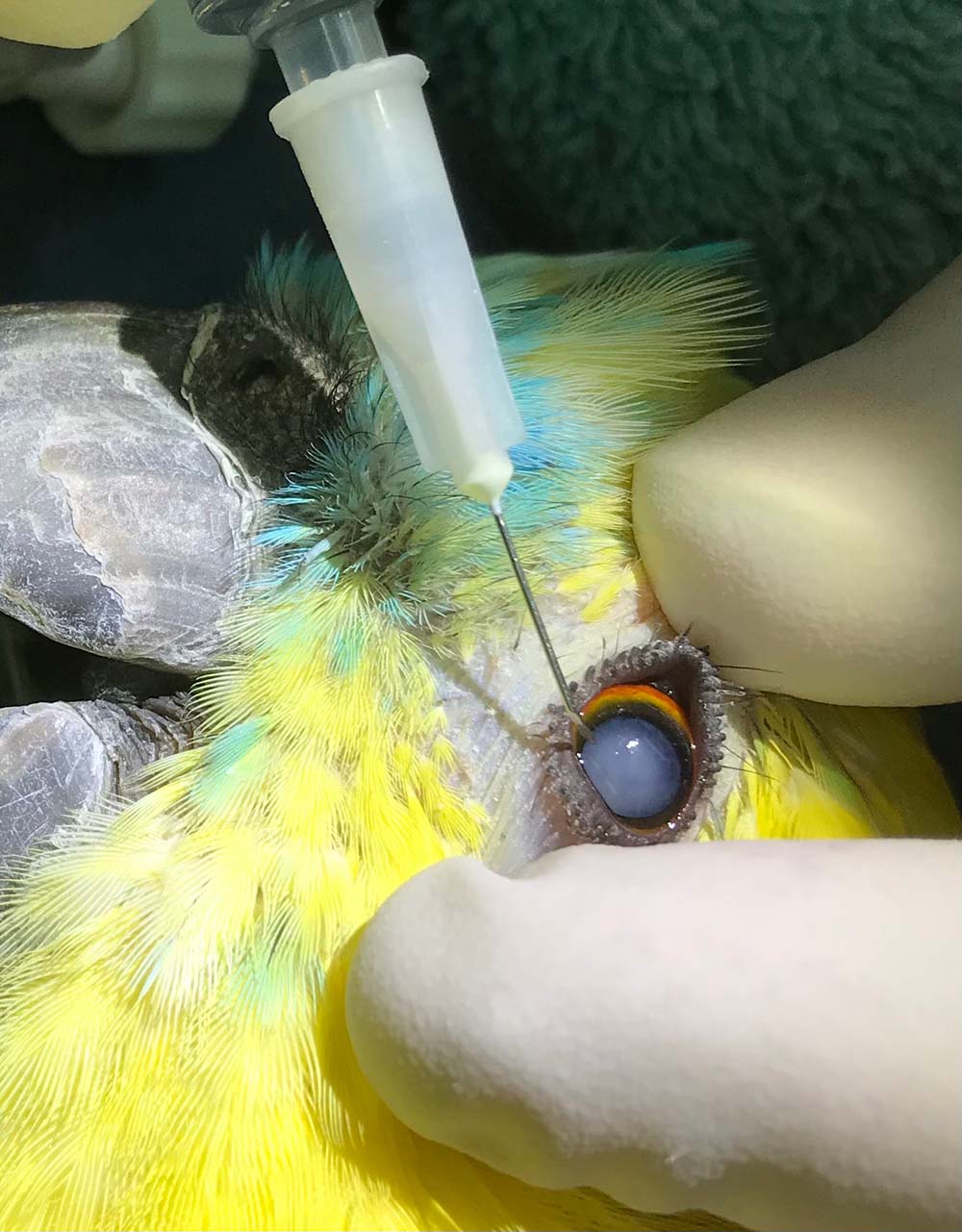
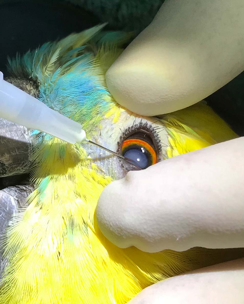
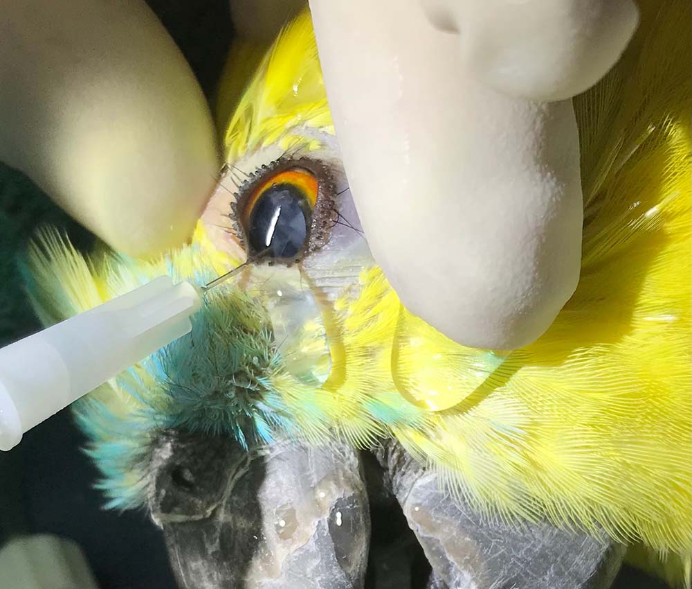
Under isoflurane anaesthesia and following dilation of the pupil using topical rocuronium, a 27G, half-inch needle was bent at the tip and used to enter the anterior chamber of the eye, incise the capsule of the lens and break down the cataract manually using a rotating action (Figures 2 to 4).
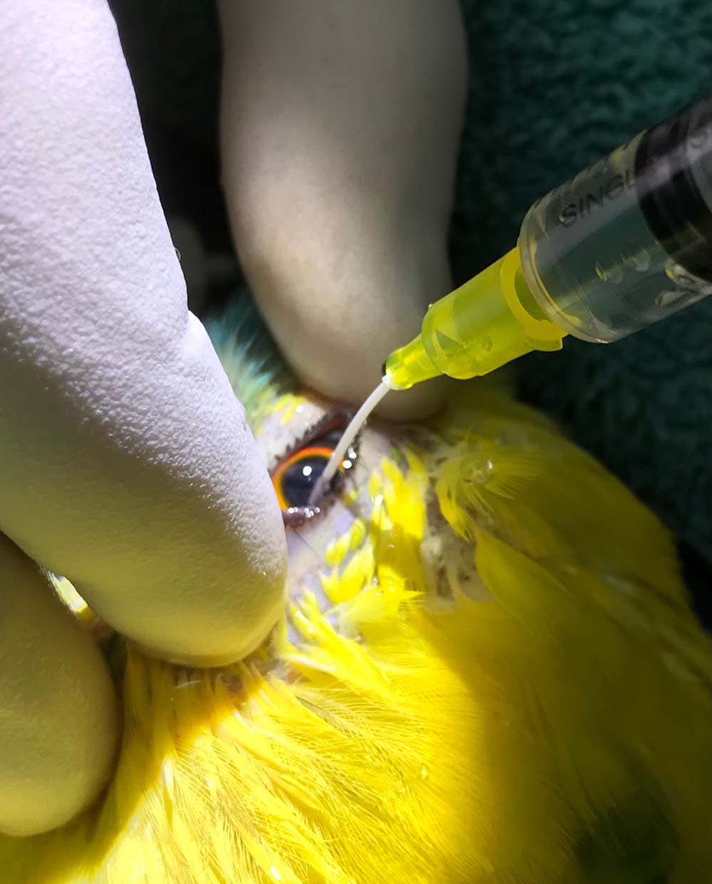
A saline-filled syringe was then attached to the needle and the cataract material gently flushed out through a lateral corneal stab incision made with a number 11 blade (Figure 5).
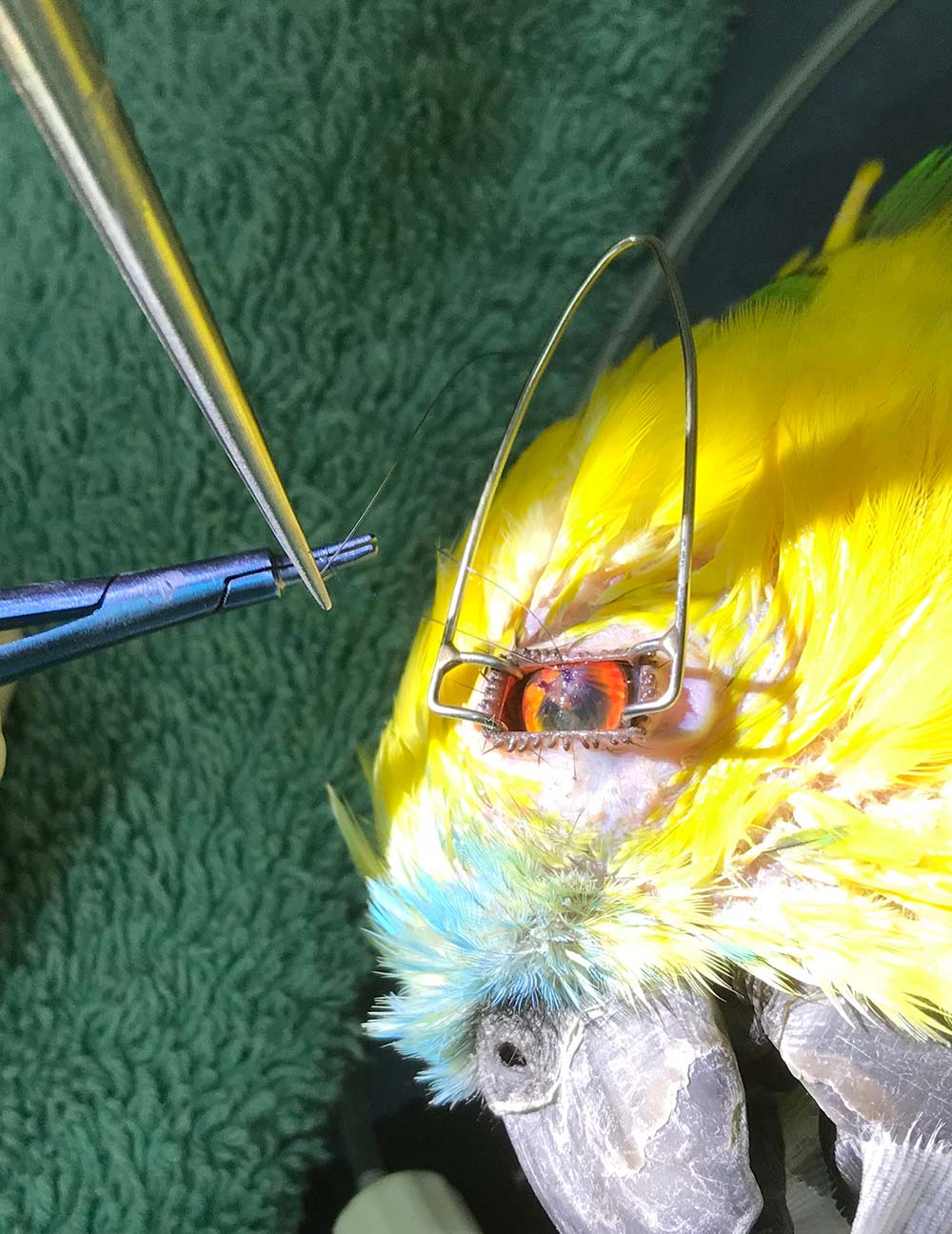
Any remaining cataract material was “flush and sucked” out using a 24G IV catheter/saline-filled syringe introduced via the lateral incision, which was then closed with two simple interrupted 6-0 polydioxanone sutures and the globe re-inflated off the needle (Figures 6 to 8).
Post-surgery – and second operation
Ten weeks following surgery, the author’s team was extremely excited that, following this relatively “low-tech” procedure, the patient was now clearly visual (Figure 9). In fact, the owner was so pleased with the results, he requested the team operate on the other eye, which thankfully also went to plan.
The patient was once again able to negotiate his aviary and interact with its flock mates, which continues to this day, now more than two years postoperatively.
Where resources are available, the author’s team will always aim to refer such cases for specialist treatment; however, under such circumstances, this procedure, which may be carried out in the general practice setting, at least offers the clinician a viable plan B and was ultimately successful in this case, with the bird now visual and once again able to enjoy a good quality of life.
- Some of the drugs in this article are used under the cascade.