14 Sept 2021
Vitamin B12 deficiency in a dog on medication for heart failure
Rory Thomson
Job Title
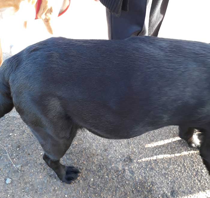
Figure 1. A male black Labrador retriever puppy called Ben.
Ben first presented at eight weeks of age for first vaccination. Ben was a male black Labrador retriever puppy and came from a reputable breeder, with a history of good elbow and hip scores of both parents. He was in good body condition and looked like a healthy puppy.
Cardiac auscultation revealed a grade II/VI heart murmur (Table 1). It was hoped it was just an innocent murmur that would be gone by second vaccination; however, the owner was advised it may be something much more significant and if it had not gone by second vaccination it should be investigated to be on the safe side.
| Table 1. The author’s “general practitioner” approach to heart murmurs in animals. It is a very simple way of scoring a murmur, which enables future monitoring. More advanced ways of describing a murmur exist in terms of when the murmur is heard in relation to systole or diastole, but that is beyond the scope of this article | |
|---|---|
| Grade of heart murmur | Definition |
| I/VI | Did I hear a heart murmur? |
| II/VI | There is a heart murmur, but it is quieter than the sound of the heart |
| III/VI | There is a heart murmur and it is the same volume as the heart |
| IV/VI | There is a heart murmur, but it is a bit louder than the sound of the heart |
| V/VI | There is a heart murmur, but it is a lot louder than the sound of the heart |
| VI/VI | I can hear the heart murmur before I put the stethoscope on the animal |
Unfortunately, by second vaccination, this heart murmur was much more audible and recorded as a grade III/VI murmur (and it was at the grade IV end of grade III). Although an excited and healthy-looking puppy, the heart rate of 172 was higher than expected. A mobile veterinary imaging company was contacted to perform echocardiography as the author did not have the equipment or expertise to perform this.
Diagnosis and treatment
The report contained various numbers and measurements that the author did not understand; however, these all came together to form a diagnosis of valvular pulmonic stenosis with a pressure gradient of 86mmHg (anything more than 80mmHg classifies as severe). Signs of right-sided remodelling and valve changes were also present.
Immediate referral for assessment by an interventional cardiologist with a view to balloon dilation was advised.
Ben was started on a beta blocker – atenolol – at 0.5mg/kg orally every 12 hours and increased to 1mg/kg to 1.5mg/kg orally over a two-week period. This was under the advice of the referral centre to slow the heart rate and reduce myocardial oxygen consumption. This was also suggested to help with accurate balloon placement and reduce the risk of some of the complications that could occur during the procedure (Figure 1).

The diagnosis of pulmonic stenosis with tricuspid dysplasia was made at the referral centre and balloon valvuloplasty was successful in reducing the pulmonic stenosis from a severe grade to a moderate grade; however, the tricuspid dysplasia was not treatable at this referral centre and the likelihood of future right-sided heart failure progression was discussed.
Following referral, Ben maintained a dose of 1mg/kg of atenolol. He was on a liquid atenolol solution, the volume of which had to be adjusted regularly as he grew. His heart murmur seemed stable at grade III/VI.
Six months old…
Approximately two months after referral, Ben presented showing signs of right-sided heart failure. At this point he was six months old, and had a grade V/VI heart murmur and palpable fluid thrill to his abdomen, indicating ascites.
Ben was a very rotund dog, but still with the same bright demeanour and waggy tail he always had. He seemed completely unaware of the severity of his condition.
At this point pimobendan was introduced at 0.2mg/kg twice daily and furosemide at 1.6mg/kg three times daily alongside his 1mg/kg atenolol.
This had the effect of causing a 20% reduction in bodyweight, which was all retained fluid. The heart murmur reduced to a grade IV/VI and the normal canine body conformation of a 6-month-old Labrador retriever was restored. Ben remained relatively stable on this treatment regimen until his postoperative check at the referral centre, 6 months after the balloon dilation (at 10 months of age).
The repeat echocardiography at this point showed the pulmonic stenosis and tricuspid dysplasia had deteriorated, causing right-sided heart failure. Ben was now at the point of either palliative care with medical management or very specialist open heart surgery for valve replacement.
The owner was not willing to put Ben through this procedure, so opted for ongoing medical management of Ben with the understanding that his lifespan was very limited. Benazepril at 0.18mg/kg once daily and spironolactone at 1.4mg/kg once daily were introduced to help the heart failure, and the atenolol weaned off slowly.
Eleven months old…
The medical management kept Ben stable until he was about 11 months old, at which point he presented again as the owners were concerned he was off his food. Ben up to this point had been a typical Labrador retriever and for him to be off his food meant something must be seriously wrong.
Ben was still his usual self on clinical exam, and seemed bright and happy as usual. His heart rate was very irregular, though, and some pulse deficits existed. The owner was not keen on any further long-distance travelling back to the original referral centre and just wanted Ben to be comfortable.
Ben was referred to a local veterinary cardiologist for an ECG to confirm the author’s suspicion of atrial fibrillation.
By the time Ben saw the cardiologist, his ascites had returned. The ECG showed some bizarre changes associated with the right-sided cardiac remodelling and confirmed the presence of atrial fibrillation. Some ventricular premature complexes were also seen and thought to be secondary to myocardial hypoxia.
Ben was started on digoxin to help with the atrial fibrillation at a dose of 4.5mcg/kg twice daily and his furosemide was increased to 2mg/kg three times daily. The owner was advised that the time for euthanasia may be getting close, particularly if no response to this treatment occurred.
The owner returned a week later and Ben had not made any significant improvement. He was still ascitic and inappetent (Figure 2). The owner was very keen to let Ben see his first birthday and wanted to try anything else to help shift the excess water to make him feel better.
Diuretic considerations
At this point Ben was already on a loop diuretic (furosemide) acting on the loop of Henle in the kidney, which was probably the most effective diuretic. He was also on a potassium-sparing diuretic (spironolactone), acting on the collection duct, although the diuretic effects of these are not as significant as their antifibrotic action. A third class of diuretics – the thiazide diuretics, which act on the distal convoluted tubule – were considered.
After comprehensive discussions with the owners about the risks of potential hypovolaemia, azotaemia and hypokalaemia, which are much higher when used alongside loop diuretics, it was decided to start hydrochlorothiazide (moduretic) at 1.8mg/kg once daily.
The plan was to try this for five days, and check biochemistry and electrolytes to ensure this was not making things worse. The potential for euthanasia if no significant improvement was seen was discussed, although the timing of euthanasia didn’t feel right, considering how bright and happy Ben seemed.
Reassessment
When reassessed five days later, Ben’s ascites had improved significantly and Ben, although not eating his usual food, was eating small amounts of a home-cooked diet and the owners were happy he was improving.
His renal parameters were at the upper end of the normal range and his electrolytes were deemed acceptable (Table 2). His clinical exam and biochemistry suggested his heart seemed stable, but he was still inappetent, which was making tablet administration difficult.
| Table 2. Biochemistry results used to check for side effects of multiple diuretics. The slight elevation of creatinine caused concern to increase hydrochlorothiazide dose any further, but were reassuring that electrolytes were normal | |
|---|---|
| Albumin | 42(25-44)g/L |
| Alkaline phosphatase | 18(20-150)U/L |
| Alanine aminotransferase | 35(10-118)U/L |
| Amylase | 796(200-1200)U/L |
| Total bilirubin | 7(2-10)umol/L |
| Blood urea nitrogen | 7.7(2.5-8.9)mmol/L |
| Calcium | 2.88(2.15-2.95)mmol/L |
| Phosphorus | 1.73(0.94-2.13)mmol/L |
| Creatinine | 136(27-124)umol/L |
| Glucose | 5.7(3.3-6.1)mmol/L |
| Sodium | 143(138-160)mmol/L |
| Potassium | 5.0(3.7-5.8)mmol/L |
| Total protein | 61(54-82)g/L |
| Globulin | 19(23-52)g/L |
Based on his biochemistry, the author was reluctant to increase the diuretics any further and wondered if anything else may help with his appetite.
Deficiency
A hypothesis was made that the amount of diuretic Ben was on, combined with his poor appetite, could be causing him to become deficient in water-soluble vitamins such as B12 as they would be being removed via increased urination quicker than they were being absorbed, and that B12 supplementation may help improve his appetite. No evidence was found in the literature that this could be the case.
Morrow et al found people showed decreases in folate concentrations on long-term diuretic therapy, yet B12 remained normal1. As these patients were not on such a high level of diuretics, the author thought it would be worth checking the B12 levels for Ben to test his hypothesis.
The author found that Ben did have low levels of B12, yet the folate levels were normal (Table 3). Ben was given 1mg hydroxycobalamin subcutaneously once weekly for six weeks.
| Table 3. These results show a normal folate (vitamin B5) and a reduced cobalamin (vitamin B12) | |
|---|---|
| Folate | 9(7.2-23.8)ng/L |
| Cobalamin | 193(240-590)ng/L |
Twelve months old…
Ben’s first birthday was halfway through this course. His appetite at this point had returned, his ascites was the best it had been in months and he even had more energy.
The B12 supplementation clearly helped Ben and improved his quality of life… unfortunately the injections did sting, and Ben started to show signs of fear and stress when coming to the surgery, rather than his usual happy self. It was decided to reduce the frequency of these injections to monthly.
Oral B12 supplementation was discussed, but the owners were keen to keep going with the injections due to the number of other oral medications that needed administering.
Fifteen months old…
Although Ben had another good, normal three months of life, he deteriorated again at about 15 months of age. This time he was no longer his happy self and seemed clinically more lethargic.
He had an irregularly irregular heart rate, but didn’t seem to have any pulse deficits and his ascites had returned.
The addition of diltiazem was considered to better control any atrial fibrillation or switching from furosemide to torasemide, but at this point it was decided the time had come for euthanasia.
Discussion
The author found this case quite interesting – particularly the response of Ben to B12 supplementation – and believes this would benefit from further research.
The approach to this case may not have been perfect and various parts of this case could be discussed, monitored and potentially managed differently, but it is a real case that has been managed for the best part within a first opinion practice, with input from various advanced practitioners and specialists where necessary.
Although closer monitoring with a veterinary cardiologist would have been beneficial in this case, the owner did not want this.
Further discussion between the author and another cardiologist revealed that the inappetence in right-sided heart failure could be due to gut oedema, which can cause a downwards spiral due to the reduced ability to absorb medication, causing a worsening of the oedema and less medication absorption. Repeated injectable diuretics for a few days could, therefore, have been a rescue therapy in this case to enable better absorption of the oral medications.
Gut oedema may also have contributed to the reduced B12 levels due to lack of absorption, meaning the injectable B12 supplementation would have been far superior to oral supplementation in this case.
The author believes a place exists for B12 supplementation in cardiac failure, and urges other practitioners to consider its use in patients with cardiac failure and inappetence. More research is needed to ascertain if this affects a significant number of cardiac patients, and to establish the mechanism of action and true benefits of supplementation.
Some drugs mentioned in this article are used under the cascade.
Acknowledgements
This case was beyond the author’s level of expertise on a number of occasions and the following veterinary surgeons had input into Ben’s care during his short life:
- Sophie Betts BVM&S, CertSAM, GPCert(Cardio), MRCVS – International Veterinary Ultrasound Society certified diagnostic small animal ultrasonographer at North East Veterinary Imaging.
- Yolanda Martinez Pereira LdaVet, CertVC, DipECVIM-CA(Cardiology), MRCVS – European and RCVS-recognised specialist in veterinary cardiology at The University of Edinburgh Royal (Dick) School of Veterinary Studies cardiopulmonary medicine department.
- Tim Bradbury BVM&S, CertVC, MRCVS – Tyne Veterinary Clinic.
- Dave Dickson DVC, MRCVS, RCVS specialist in veterinary cardiology – HeartVets.
It is important for all vets to be aware of their limitations and not be scared to ask for help when needed.
References
- Morrow LE and Grimsley EW (1999). Long-term diuretic therapy in hypertensive patients: effects on serum homocysteine, vitamin B6, vitamin B12, and red blood cell folate concentrations, Southern Medical Journal 92(9): 866-870.